• Log In

Microvascular Therapeutics and Imaging Laboratory

Department of Biomedical Engineering and Beckman Laser Institute, University of California, Irvine

  • Home
    • News
    • Archived News
  • Research
    • Optical Imaging Instrumentation Development
    • in vivo Monitoring of Microvascular Response to Novel Therapeutics
    • Real-time Non-invasive Blood Flow Imaging
    • Engineering of Tissue Optical Properties
  • Publications
  • Members
  • Lab Openings
  • Software
  • Presentations
  • Resources
  • Links
  • Lab Intranet

Real-time Non-invasive Blood Flow Imaging

The current port wine stain (PWS) treatment method is based on the premise that acute photocoagulation of the targeted microvasculature results ultimately in replacement of the damaged blood vessels with normal-sized ones. Unfortunately, the ultimate course of treatment to achieve this goal is primarily determined by the physician based on their clinical experience. We postulate that regions of persistent perfusion exist during and after treatment. These regions are largely “invisible” to the treating physician due to the current lack of appropriate clinical instrumentation. We suspect this issue contributes to the poor efficacy of the current treatment method. Immediate retreatment of these regions may result in an improved treatment outcome per session, reducing the number of treatment sessions required for complete PWS blanching.

Figure 1. Schematic of cart based clinical LSI instrument

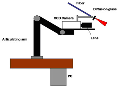
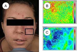
To address this need, we propose use of laser speckle imaging (LSI) to provide real-time, quantitative feedback during laser surgery. We have collected preliminary patient data using our cart-based laser speckle imaging (LSI) instrument (Fig. 1). Raw speckle images were acquired from a PWS skin predefined site enclosed within the black line (Fig. 2A) prior to and 15 minutes after laser therapy. Prior to treatment (Fig. 2B), the blood flow distribution was fairly heterogeneous. After treatment (Fig. 2C), blood flow decreased presumably due to PWS blood vessel photocoagulation. Collectively, these data confirmed that regions of persistent perfusion remain immediately after laser therapy.

For more information, please refer to our recent publication in Lasers in Surgery and Medicine.

clinical_2
Figure 2. (A) 24-bit RGB and speckle flow index images computed from raw speckle images (B) prior to and (C) 15 min after laser therapy by a highly experienced physician

Collaborators: Kristen Kelly, Stuart Nelson, UC Irvine

Recent Posts

  • Mio Jiang awarded a summer research fellowship
  • Poster presented by Aarti Dachepalli at the UCI UROP symposium
  • New review paper by Dr. Christian Crouzet published in Neurophotonics on wide-field functional imaging of the brain
  • Danny Xie will give a virtual poster presentation at the 2022 International Stroke Conference
  • New NSF Graduate Research Fellow in MTI lab

Recent Comments

  • Anonymous on Hello world!

Archives

  • June 2023
  • May 2023
  • February 2022
  • April 2020
  • June 2019
  • May 2019
  • February 2019
  • June 2018
  • May 2018
  • June 2017
  • October 2016
  • September 2016
  • August 2016
  • July 2016
  • December 2015
  • October 2015
  • September 2015
  • May 2015
  • April 2015
  • September 2014
  • July 2014
  • June 2014
  • May 2014
  • March 2014
  • February 2014
  • January 2014
  • October 2013
  • September 2013

Categories

  • Archived News
  • Conference Presentations
  • News
  • Publications
  • Uncategorized
  • UROP

Copyright © 2025 · Beautiful Pro Theme on Genesis Framework · WordPress · Log in